

Web コンテンツ・ビューアー
アクション
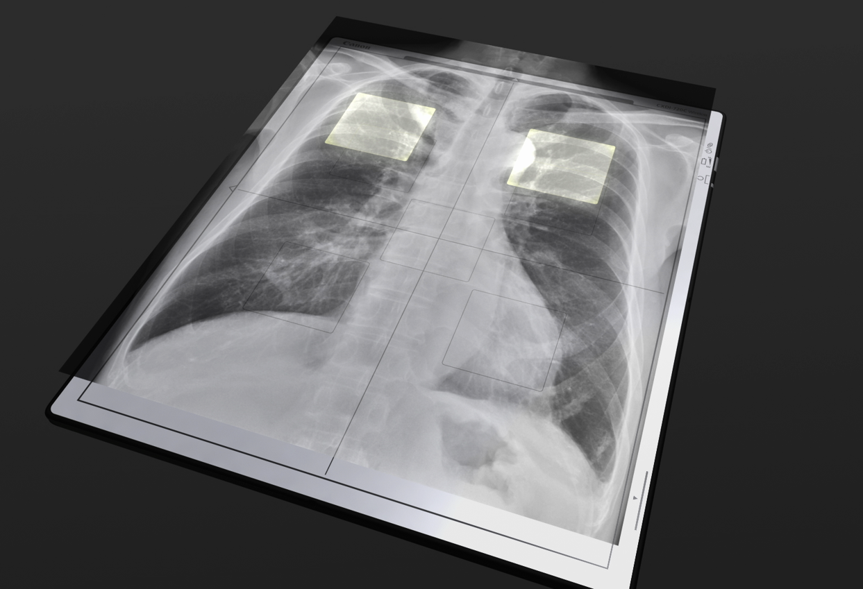

For the Ideal AEC Radiography
The CXDI Elite series, the world’s first built-in AEC FPD, allows for automatically terminated exposures without the use of an additional receptor (ion chamber, solid state paddle, etc.).
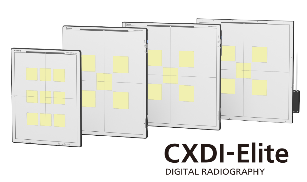
CXDI-Elite Assists Auto Exposure Control
Built-in AEC Assistance (BiAA) technology
enables the detector to monitor the accumulated
pixel values in real time at each AEC region of
interest (ROI) and signals the generator to stop
exposure once a preset value is reached.
There
are 5 or 9 AEC Regions of Interest (ROI) depending
upon model.
Compatible Detectors: CXDI-720C
Wireless, CXDI-820C Wireless, CXDI-420C Wireless,
CXDI-420C Fixed
*Option software sold separately and Multibox (MB-02) is also required
Expanding AEC to new applications
- No need for traditional AEC receptor (ion chamber, solid state paddle, etc.)
- Built-in AEC Assistance works via both wireless and wired communication.
- Radiation dose can be optimized, even in free-position imaging at the bedside, tabletop, wheelchairs, and gurneys.

Dose Control Performance
- Phantom Study: Exposure Index (EI) Stability Across Tube Voltage and Patient Thickness
Using PMMA (acrylic) phantoms, this
study evaluated the stability of the Exposure Index (EI) in
BiAA wireless detectors under varying tube voltage (kV) and
patient thickness conditions, in comparison with
conventional phototimers.
Across a wide range of imaging
settings, BiAA consistently maintained normalized EI values
close to 1.0, demonstrating improved stability compared with
conventional phototimers. These results highlight BiAA’s
potential to deliver consistent image quality and reliable
dose monitoring across different clinical scenarios.
This
linear response will negate the need for compensation
factors in the calibration of the AEC. Thus resulting in
more reliable imaging with simpler service setup.
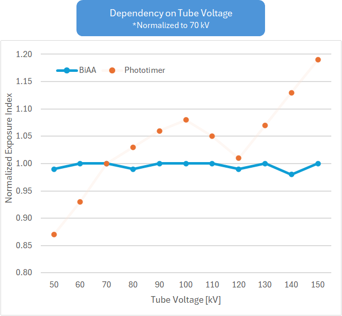
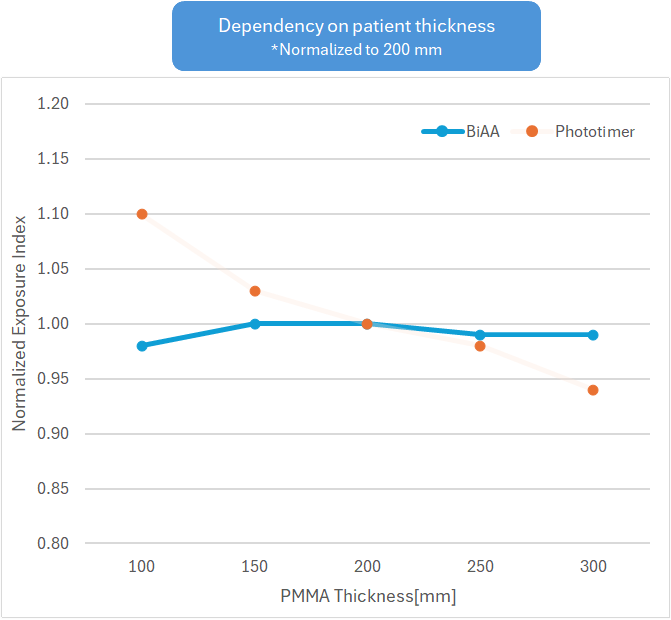
Excerpted from Toyonaga et al., Kyoto University Hospital, 1st Japanese Congress of Radiological Technology and Medicine (2024)